Simplified in Practice: ECG features of posterior MI
M3 India Newsdesk Sep 16, 2021
In this part of our series of Simplified in Practice, the focus topic is posterior myocardial infarction and its importance in clinical practice. Posterior myocardial infarction generally has always had a tendency of being overlooked. For medical practitioners, it is crucial to be alert for signs of posterior MI for any development of further complications.
A posterior infarction occurs in 15-20% of STEMIs, most often in conjunction with an inferior or lateral infarction. Isolated posterior MI is a less frequent occurrence (3-11 per cent of infarcts). Posterior expansion of an inferior or lateral infarct results in a much greater region of myocardial damage, increasing the likelihood of left ventricular failure and mortality.
Isolated posterior infarction necessitates urgent coronary reperfusion therapy. However, due to the absence of apparent ST elevation in this disease, the diagnosis is often overlooked. In any patient with an inferior or lateral STEMI, be alert for signs of posterior MI.
How to determine whether a patient has had a posterior infarction?
Due to the fact that the conventional 12-lead ECG cannot directly see the posterior myocardium, reciprocal alterations indicative of STEMI are sought in the anteroseptal leads V1-3.
The following alterations in V1-3 indicate a posterior MI:
- ST depression horizontally
- R waves that are tall and wide (>30 ms)
- T waves that are upright
- Dominant R wave (R/S ratio greater than one) in version 2
Horizontal ST depression in the anteroseptal leads (V1-3) might trigger suspicion of posterior MI in individuals who present with ischaemic symptoms.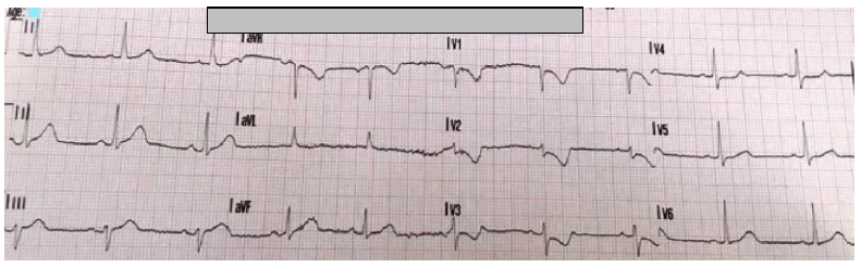 The presence of ST-elevation and Q waves in the posterior leads confirms posterior infarction (V7-9).
ECG abnormalities in V1-3
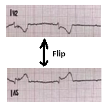
The anteroseptal leads go from the anterior precordium to the posterior myocardium's internal surface. Due to the fact that posterior electrical activity is recorded from the anterior side of the heart, the characteristic damage pattern of ST-elevation and Q waves is inverted:
- The ST-elevation transforms into ST depression.
- Q waves morph into R waves.
- Inversion of the terminal T wave results in the formation of an upright T wave.
- The gradual development of pathogenic R waves (the "Q wave equivalent") in posterior infarction parallels the progression of Q waves in anteroseptal STEMI.
This diagram depicts the reciprocal connection between the ECG abnormalities associated with STEMI and those associated with posterior infarction. The preceding picture has been flipped to show a posterior infarction in V2. As you can see, the ECG now resembles that of a classic STEMI.
Leads in the posterior
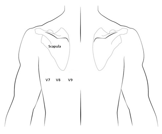
Leads V7-9 are inserted in the following locations on the posterior chest wall:
- V7 - Left posterior axillary line, parallel to V6
- V8 - In the same horizontal plane as V6, the tip of the left scapula
- V9 - In the same horizontal plane as V6, the left paraspinal region
- Leads in the posterior ECG location in V7 V8 V9
- Lead implantation in the posterior V7, V8, and V9
ST-elevation is usually mild in V7-9 — remember that just 0.5 mm of ST elevation is needed to diagnose posterior MI.
This is part five of our new series- Simplified in Practice where we break down ECG interpretation for various conditions. To read part one and two of the series, click here: Simplified in Practice: ECG basics & how to interpret, Simplified in Practice: How to detect myocardial ischaemia?, Simplified in Practice: Anterior Myocardial Infarction- Clinical nuggets on ECG characteristics & Simplified in Practice: What are the ECG changes when inferior STEMI starts?
Click here to see references
Disclaimer- The views and opinions expressed in this article are those of the author's and do not necessarily reflect the official policy or position of M3 India.
The author is a practising super specialist from New Delhi.
-
Exclusive Write-ups & Webinars by KOLs
-
 Daily Quiz by specialty
 -
 Paid Market Research Surveys
 -
Case discussions, News & Journals' summaries
