Simplified in Practice: What are the ECG changes when inferior STEMI starts?
M3 India Newsdesk Aug 26, 2021
This article on inferior wall myocardial infarction (IWMI) is the next part of our series- Simplified in Practice, and it throws light on on the diagnostic evaluations made through ECGs- specifically indications of RCA obstruction and ECG changes that occur when inferior STEMI begins.
ECG nuggets - Inferior wall myocardial infarction
40% to 50% of all myocardial infarctions (MIs) occur in the inferior wall. Although it has a better prognosis than anterior myocardial infarction (in-hospital mortality is just 2 to 9%), some concomitant characteristics suggest a poor result.
Diagnostic evaluation for electrocardiograms:
- Elevation of the ST leads in leads II, III, and aVF
- These alterations may be preceded by hyperacute T waves
- Reciprocal ST depression in the aVL
- Progression of Q waves in the II, III, and aVF
The following characteristics are associated with a poor prognosis:
- 40% of patients have concurrent right ventricular infarction and this subset of patients suffer severe hypotension in response to nitrates
- 20% of the patients have severe bradycardia owing to 2nd or 3rd degree AV block
- Inferior wall ischaemia extending to posterior wall infarction also carries poor prognosis
Keep aVL in mind
- As it is the sole lead facing the superior portion of the ventricle, aVL is the only lead that is genuinely reciprocal to the inferior wall. As a result, it is a sensitive indicator of inferior infarction.
- ST depression in aVL has been found to be more common than STE in inferior leads in patient cohorts with inferior occlusion myocardial infarction (OMI).
- ST depression in the aVL is seen in 91% of “subtle” inferior STEMIs that do not satisfy STEMI criteria but exhibit occlusion on PCI.
Which artery is the source of the problem?
STEMI in the inferior coronary artery may occur as a consequence of a blockage of any of the three major coronary arteries:
- In 80% of instances, the right coronary artery (RCA) is dominant and in 18% of cases, the left circumflex artery (LCx) is dominant.
- A “type III” or “wraparound” LAD may sometimes cause a unique pattern of inferior and anterior ST elevation.
Although both RCA and LCx occlusions may result in inferior wall infarction, the exact region of infarction and therefore the ECG pattern is somewhat different in each case. The RCA area encompasses the inferior wall's medial portion, including the inferior septum. In RCA occlusion, the injury current is channelled inferiorly and rightward, resulting in ST elevation in lead III > lead II (as lead III is more rightward facing).
The LCx region encompasses the inferior wall's lateral portion and the left posterobasal section. In LCx blockage, the injury current is channelled inferiorly and leftward, resulting in ST elevation in lateral leads I and V5-6. These distinctions enable electrocardiographic discrimination between RCA and LCx occlusions.
Indications of the RCA obstruction
- ST elevation in lead III > lead II
- Reciprocal ST depression in lead I
- Right ventricular infarction signs: STE in V1 and V4R
The blockage of the circumflex artery is indicated by:
- A rise in the ST in lead II equals a rise in the ST in lead III
- No reciprocal ST depression in lead I
- Signs of lateral infarction: ST elevation in lateral leads I and aVL or V5-6
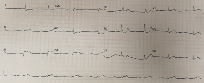
ECG changes when inferior STEMI starts
- Early ST elevation and Q-wave development in lead III
- Hyperacute (peaked) T waves in leads II, III, and aVF with a loss of relative R wave height
- Reciprocal ST depression and T wave inversion in aVL
- ST elevation in lead III > lead II is compatible with an RCA blockage, the slight ST elevation in V4R is also consistent

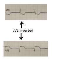
- Take note of how the shape of the ST segment in aVL is an identical mirror copy of that of lead III. The idea of reciprocal change may be shown further by inverting lead aVL. Take note of how the ST morphology now resembles that of lead III:
Inferior STEMI patients with atrioventricular block and bradycardia
Second or third-degree AV block may occur in up to 20% of patients with inferior STEMI. There are two possible explanations for this:
- Ischaemia of the AV node as a result of decreased blood flow via the AV nodal artery. This artery originates from the RCA in about 80% of cases, which explains its involvement in inferior STEMI caused by RCA blockage.
- The Bezold-Jarisch reflex refers to an increase in vagal tone as a result of ischaemia.
In 50% of instances, the conduction block develops as a step-by-step development from 1st-degree heart block through Wenckebach to full heart block, or as a sudden start of second or third-degree heart block. Additionally, patients may exhibit symptoms consistent with sinus node dysfunction, including sinus bradycardia, sinus pauses, sinoatrial exit block, and sinus arrest.
As with AV node dysfunction, this may be caused by increased vagal tone or SA node ischaemia (the SA nodal artery is supplied by the RCA in 60 per cent of people).
This is part four of our new series- Simplified in Practice where we break down ECG interpretation for various conditions. To read part one and two of the series, click here: Simplified in Practice: ECG basics & how to interpret, Simplified in Practice: How to detect myocardial ischaemia? & Simplified in Practice: Anterior Myocardial Infarction- Clinical nuggets on ECG characteristics
Click here to see references
Disclaimer- The views and opinions expressed in this article are those of the author's and do not necessarily reflect the official policy or position of M3 India.
The author is a practising super specialist from New Delhi.
-
Exclusive Write-ups & Webinars by KOLs
-
 Daily Quiz by specialty
 -
 Paid Market Research Surveys
 -
Case discussions, News & Journals' summaries
