Familial Adenomatous Polyposis Management
M3 India Newsdesk Jan 17, 2024
The article discusses the clinical presentation, diagnostic approaches, and treatment options for FAP. It also emphasises the importance of genetic testing and the challenges posed by limited resources.
Familial Adenomatous Polyposis (FAP)
Case study 1
The 14-year-old male presented with a history of bright red rectal bleeding, intermittent lower abdominal pain, occasional diarrhoea, and sporadic fever persisting for the past 1.5 years.
Notably, there's a family history of premature deaths; his father passed away at 36 years old, and his elder sister at 14 years old, both exhibiting similar symptoms of rectal bleeding, yet remaining undiagnosed.
Examination
- During examination, the patient appeared pale but was stable in terms of temperature and hemodynamics.
- Abdominal examination revealed softness, absence of tenderness, and no distension.
- Other systemic examinations, including fundoscopy, did not reveal any significant findings.
- Blood tests indicated normal blood counts but showed signs of anaemia (Hemoglobin-8.0 g/dl, PCV-26 g %).
- Other blood investigations, including tumour markers (CEA and CA-19.9), fell within normal ranges.
(A)

(B)
(C)
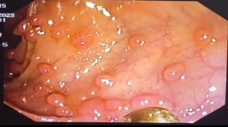
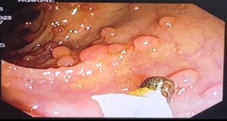
Fig. A – Pedunculate polyp
Fig. B and C show a Colonoscopic picture of both sessile and pedunculated Polyp
- A colonoscopy revealed a concerning finding of multiple pedunculated and sessile colonic polyps, exceeding 100 in number, spanning from the rectum to the cecum.
- Biopsy results from a rectal polyp indicated findings consistent with focal active colitis.
- A CT scan of the abdomen and pelvis disclosed multiple heterogeneously enhancing polypoidal endophytic soft tissue densities attached to the bowel walls in various segments including the rectum, sigmoid colon, descending colon, mid aspect of the transverse colon, and ascending colon.
- The patient underwent a laparoscopic total proctocolectomy in two stages. Initially, a laparoscopic procedure was used to mobilise the caecum, ascending colon, transverse colon, descending colon, sigmoid colon, and rectum, with the ligation of associated vessels such as the left colic and right colic artery.
- In the subsequent phase, a lower midline incision was made to resect the mobilised large intestine with the rectum, and an ileoileal pouch was constructed.
- The Soave procedure was employed to pull through the ileal pouch, followed by the creation of a loop ileostomy. The skilled gastrointestinal surgical team at the Rajiv Gandhi Government General Hospital conducted the ileal anal pouch anastomosis with loop ileostomy, which the patient tolerated well.
- Upon examination of the resected specimen, a significant finding was noted: multiple pedunculated polyps, exceeding 550 in number, were observed throughout the colon.
- Microscopic analysis of these polyps revealed cystically dilated glands with the presence of neutrophils infiltrating the lumens. Moreover, focal dysplasia was identified within the glands, characterised by nuclear stratification, hyperchromatic nuclei, and loss of apical mucin.
- The histopathological findings align with a condition associated with the development of numerous colonic polyps, possibly indicative of a hereditary polyposis syndrome.
- However, due to limitations in the facility's resources and financial constraints affecting the patient and high-risk family members, genetic testing, which could potentially provide critical insights into the underlying genetic cause of this condition, was not conducted.
- It's positive to note that the post-operative phase proceeded without complications, and the patient has made a successful recovery.
- At six months of follow-up, the patient reports no complaints and is currently free from disease.
Discussion
Introduction
Familial adenomatous polyposis (FAP) is a genetic disorder primarily characterised by the presence of numerous colorectal adenomatous polyps, usually exceeding a count of 100.
Epidemiology
- Familial adenomatous polyposis (FAP) is estimated to affect approximately 1 in 8000 to 1 in 18,000 individuals and represents less than 1 per cent of all colorectal cancers in the United States.
- This condition is observed with equal frequency in both sexes and exhibits a global distribution, affecting populations worldwide.
Genetics
- Familial adenomatous polyposis (FAP) and its variants are primarily caused by mutations within the Adenomatous Polyposis Coli (APC) gene, which functions as a tumour suppressor and is located on chromosome 5q21-q22.
- The inheritance pattern of FAP is autosomal dominant, typically exhibiting nearly complete penetrance concerning colonic polyposis.
- Approximately 25 per cent of FAP cases arise from de novo mutations in the APC gene, occurring in individuals without a family history of FAP.
- Despite lacking a familial background, these individuals can pass the mutation to their offspring, thereby perpetuating the genetic risk.
Clinical manifestations
- Typically, many individuals affected by FAP remain asymptomatic until the disease progresses or complications arise, often presenting with symptoms indicative of colorectal cancer.
- Common symptoms that may prompt investigation or diagnosis include gastrointestinal bleeding, abdominal pain, and diarrhoea.
Familial adenomatous polyposis (FAP) exhibits a wide array of manifestations, predominantly involving the colon but also extending to various extracolonic sites:
Classic FAP
- Characterised by the presence of hundreds to thousands of adenomatous colorectal polyps.
- These polyps tend to appear during the second or third decade of life, with onset noted as early as eight years old.
- If left untreated, colorectal cancer develops in nearly all cases, typically diagnosed around age 39.
Attenuated FAP (AFAP)
- A milder form characterised by fewer adenomas (10 to 99), diagnosed at a later age (mean ages of 44 for adenomas and 58 for cancer).
- The distribution of polyps tends to be more proximal in the colon.
- While the overall risk of colorectal cancer in AFAP is lower than in classic FAP.
- Extracolonic manifestations: FAP involves various extracolonic manifestations affecting different organ systems:
Gardner syndrome
- This includes desmoid tumours, cysts, lipomas, osteomas, and fibromas, among others.
- These manifestations are essentially interchangeable with FAP.
Turcot syndrome
- Historically associated with familial colon cancer and brain tumours, primarily medulloblastomas and gliomas.
- This term has also been used in Lynch syndrome cases with brain tumours.
Gastric polyps
- Most FAP patients have fundic gland polyps, small sessile polyps in the stomach body.
- Gastric adenomas, though less common, carry a low but definite risk of progression to cancer.
Gastric adenocarcinoma and proximal polyposis of the stomach (GAPPS):
A rare variant linked to specific mutations in the APC gene, characterised by >100 polyps in the gastric body and fundus with antral sparing, often associated with a high risk of gastric cancer but without significant colon polyposis.
Duodenal adenomas
Common in FAP (34 to 90 per cent), particularly in the ampullary and periampullary regions, with a 3.1 to 5 per cent lifetime risk of duodenal cancer.
Desmoid tumours
Mesenchymal tumours occur in 10 to 15 per cent of FAP patients, commonly in the abdomen, causing potential complications due to their location.
Other manifestations
Benign thyroid nodules and thyroid cancer, hepatoblastomas, brain tumours like medulloblastoma, and various other manifestations such as sebaceous or epidermoid cysts, lipomas, osteomas, fibromas, dental abnormalities, juvenile nasopharyngeal angiofibroma, adrenal adenomas, and congenital hypertrophy of the retinal pigment epithelium (CHRPE).
Laboratory features
- In familial adenomatous polyposis (FAP), most individuals do not show abnormal laboratory findings.
- However, some patients may exhibit signs of iron deficiency anaemia.
- This anaemia may result from unrecognised bleeding stemming from the adenomas in the colon.
Diagnosis
Clinical suspicion
FAP should be considered in individuals found to have 10 or more cumulative colorectal adenomas.
Even if the absolute number of adenomas is lower, suspicion of FAP might arise if there is a history of adenomas coupled with extracolonic features like duodenal/ampullary adenomas, desmoid tumours, papillary thyroid cancer, congenital hypertrophy of the retinal pigment epithelium (CHRPE), epidermal cysts, or osteomas.
Genetic testing
- Genetic testing for FAP and MUTYH-associated polyposis (MAP) is recommended in individuals with 10 or more adenomas due to overlapping clinical features between these conditions.
- Genetic testing might involve an initial panel that includes genes associated with polyposis, such as APC and MUTYH, and in some cases, broader multigene panels encompassing other relevant genes like DNA polymerase ε (POLE), δ (POLD1), and Gremlin 1 homolog (GREM1).
Genetic counseling
Before genetic testing, individuals should receive genetic counselling to understand the implications and potential outcomes of the test, especially considering familial and hereditary aspects.
Family evaluation
- Identification of an APC mutation in an individual with suspected or confirmed FAP prompts further evaluation of at-risk relatives.
- First-degree relatives should be offered genetic testing.
- Additionally, consideration might be given to second-degree relatives, particularly in cases where a family member declines testing or has passed away.
- Genetic evaluation in minors, generally around 10 to 12 years of age, can precede screening colonoscopy.
- Earlier testing might be considered in families with a history of early-onset tumours, such as childhood hepatoblastoma.
Prenatal testing and preimplantation genetic diagnosis (PGD):
These options allow prospective parents to make informed decisions regarding their family planning while considering the risk of transmitting the condition to their offspring.
Differential diagnosis
Several conditions share similarities in presenting with multiple adenomatous polyps, including:
MUTYH-associated polyposis (MAP)
- This condition, typically autosomal recessive, is caused by mutations in the MUTYH gene.
- Individuals with MAP may exhibit multiple colonic adenomas and sometimes duodenal adenomas.
- Distinguishing MAP from FAP requires genetic testing.
Polymerase proofreading-associated polyposis (PPAP)
Caused by mutations in DNA polymerase ε (POLE) and δ (POLD1), PPAP can present with multiple colonic adenomas and may need differentiation through genetic testing.
Gremlin 1 homolog (GREM1) duplication
Reported cases with GREM1 duplication have shown multiple colonic adenomas, duodenal adenomas, and desmoid tumours.
Attenuated familial adenomatous polyposis (AFAP)
- Individuals with AFAP may have relatively fewer colorectal adenomas, making clinical distinction from conditions like Lynch syndrome or sporadic adenomas challenging.
- Genetic testing is essential for a definitive diagnosis.
Differentiation among these conditions—FAP, MAP, PPAP, AFAP, Lynch syndrome, or sporadic adenomas—often relies on Genetic testing due to overlapping clinical features.
Treatment
Colectomy is a recommended treatment for patients diagnosed with classic Familial Adenomatous Polyposis (FAP) due to the nearly 100 per cent risk of developing colorectal cancer (CRC) in untreated individuals.
Indications for colectomy in FAP
- Documented or suspected colorectal cancer
- Severe symptoms due to colonic neoplasia (such as gastrointestinal bleeding)
- Adenomas with high-grade dysplasia or multiple adenomas larger than 6 mm
- Significant increases in polyp number over consecutive examinations
- Inability to adequately survey the colon due to numerous diminutive polyps
Extent of colon resection
Surgical options for FAP patients include
- Total proctocolectomy with end-ileostomy
- Total proctocolectomy with ileal pouch-anal anastomosis (IPAA)
- Total colectomy with ileo-rectal anastomosis (IRA)
Timing of colectomy
- Urgent or semi-elective colectomy is recommended for documented or suspected colorectal cancer or adenomas with high-grade dysplasia.
- Early colectomy, near the time of diagnosis, is advised for symptomatic patients or those with multiple 6 to 10-mm polyps that cannot be cleared endoscopically.
- Elective colectomy might be delayed until the late teens or early twenties for classic FAP patients with sparse (<10) or small (<5 mm) adenomas.
Surveillance post-colectomy
- Colectomy does not eliminate the risk of cancer as tumours may develop in the rectum (in IRA patients), the anal transition zone, or within the ileal pouch (IPAA patients).
- Regular surveillance with endoscopic evaluations of the rectum or ileal pouch every 6 to 12 months is recommended to detect any adenomas or cancers developing in these areas.
- Patients who have undergone total proctocolectomy with IRA or IPAA remain at risk for adenomas and adenocarcinoma in the ileal pouch, though the risk is comparatively lower than in the native colon.
- Pouchitis may occur in a subset of patients following IPAA surgery but tends to be less severe than in individuals with inflammatory bowel disease.
Chemoprevention
Aspirin and Nonsteroidal Anti-Inflammatory Drugs (NSAIDs)
- Sulindac, an NSAID, has been used to reduce rectal polyp burden in some cases after surgery and during endoscopic surveillance.
- However, its capacity to prevent colorectal cancer development is unclear.
- Aspirin and resistant starch did not significantly reduce polyp number or size in a randomised trial involving patients with FAP.
Erlotinib
This epidermal growth factor inhibitor has shown promise in reducing duodenal and colorectal polyp burden in patients with FAP when used alongside sulindac. However, the long-term benefits and side effects of erlotinib require further investigation.
COX-2 Inhibitors
Celecoxib, a COX-2 inhibitor, has shown a modest reduction in colonic and duodenal adenomas. However, its use has been limited due to increased cardiovascular risks associated with this class of drugs.
Eflornithine
- An inhibitor of ornithine decarboxylase, eflornithine, has been studied in combination with sulindac or alone.
- The results did not significantly reduce the rates of disease progression, although there were indications of potential benefit in patients with an intact colon.
Curcumin
- Curcumin, a compound derived from turmeric, showed promise in reducing adenoma numbers and sizes when combined with quercetin in a small series of FAP patients.
- However, a randomised trial evaluating curcumin alone did not show a significant difference in the mean number or size of polyps between the treatment and placebo groups.
The role of these chemopreventive agents in preventing colorectal cancer or reducing adenoma burden in FAP patients requires more extensive and controlled studies to establish their efficacy, safety, and long-term effects before considering them as standard interventions in clinical practice.
Disclaimer- The views and opinions expressed in this article are those of the author and do not necessarily reflect the official policy or position of M3 India.
About the author of this article: Dr Jimmy Patel is a practising gastroenterologist in Chennai.
-
Exclusive Write-ups & Webinars by KOLs
-
 Daily Quiz by specialty
 -
 Paid Market Research Surveys
 -
Case discussions, News & Journals' summaries
