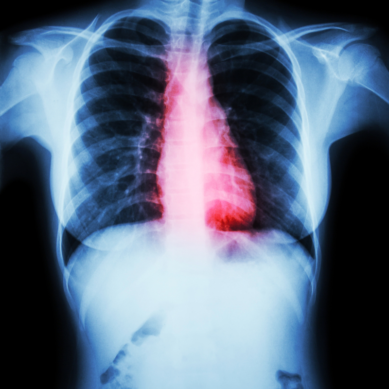
Seeing the bigger picture—the use of imaging in congenital heart disease
EuroEcho 2019 Congress News Dec 17, 2019
“There have been some really interesting presentations and sessions on imaging in congenital heart disease at EuroEcho 2019,” says Doctor Tara Bharucha (University Hospitals Southampton NHS Foundation Trust, Southampton, UK), EACVI ex officio Board member representing ‘Congenital Heart Disease’.
“From teaching courses, clinical cases and posters to specialised symposia and the Joint Session with the American Society of Echocardiography on predicting right ventricular failure with different congenital defects, which was particularly informative.”
“As in many areas of cardiology, imaging plays a very important role in the management of congenital heart disease, but its function can often be different in congenitally malformed hearts compared with acquired cardiology, partly due to anatomical anomalies and partly because of haemodynamic challenges.”
“We regularly see complex lesions that are unique—defects can be categorised but, within each specific diagnosis, there is wide variation. Imaging is essential to help individualise the management approach.”
Regarding the imaging techniques used, Dr. Bharucha is keen to mention that while echocardiography is still the primary modality for imaging congenital heart disease, other techniques are being increasingly used due to technological advances. “There is greater uptake of computed tomography (CT) and cardiovascular magnetic resonance (CMR) than in previous years,” she explains. “These modalities are much more accessible—the radiation dose with CT is acceptably low for use in small babies and the faster speed of CMR sequences means that a general aesthetic is not always necessary for children.”
And what new advances lie ahead? “3-D printing of congenital heart disease, using CT scans, is gaining a lot of attention at the moment,” says Dr. Bharucha. “3-D printing allows us to visualise individual nuances that are needed for optimal surgical planning. The 3-D printed models are also useful for informing parents and for teaching.” She continues, “Computer modelling with machine learning and artificial intelligence is also being explored to help individually tailor surgery. Further, these tools can help predict how surgical repair will progress as the child grows—computer simulation of hypothetical growth and the effect on blood flow is a new and interesting area.”
This article is a news release from EuroEcho 2019 Congress Meeting. Read the original here.
-
Exclusive Write-ups & Webinars by KOLs
-
 Daily Quiz by specialty
 -
 Paid Market Research Surveys
 -
Case discussions, News & Journals' summaries
