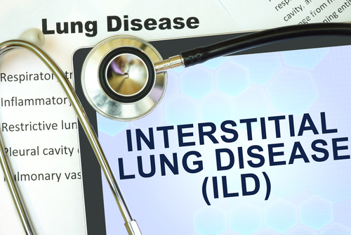
IIT Kharagpur researchers develop tools to detect interstitial lung diseases and lung cancer
IANS Dec 06, 2018
Researchers of IIT Kharagpur have developed a decision support system to diagnose malignant lung tissues and other interstitial lung diseases (ILD), said an official statement on December 5.
"Biopsy especially in the lungs is a critical process, hence conducted only after initial medical analysis is done by expert radiologists. The developed system uses non-invasive and comparatively affordable methods of image analysis that would aid the radiologists to identify malignancy by reading growth in the lung nodules.
"The other system will help identify interstitial disease patterns in HRCT images depicting the lung tissue texture," lead researcher Sudipta Mukhopadhyay explained. The malignancy detecting tool detects the lung nodule, segments the nodule, and provides a way to modify segmentation, retrieve similar nodules from the database with their report and assess the chance of malignancy of the query nodule based on the retrieval results, the statement added.
"The novelty of the system lies in its India-centric reference point i.e. the medical image scan database used for reference is sought from the Indian patient population," researcher Shrikant Mehre said. Mehre mentioned that they worked with Niranjan Khandelwal and his team from PGIMER Chandigarh for data ground truth and clinical data. Also, a foreign database such as LIDC-IDRI and Med GIFT ILD database have been used. The biopsy cases were primarily taken from PGIMER.
"The ILD tool is developed by incorporating feedback from expert radiologists to make it easy to use for non-tech savvy clinicians. The software is equipped with necessary modules such as automatic segmentation of lung boundary and pathological region within lung area, provision to modify the boundary, retrieving similar segments from the database with their report and assess the probability of the pathological segment to be a particular ILD category based on the retrieval results," the statement said.
The mapping of disease is performed by doctors based on the ILD pattern and clinical inputs. "We have successfully tested both software systems at AIIMS Delhi. Ashu Seith Bhalla and her team provided the neutral test site required for the validation. Currently, our success rate is higher than 80% in identifying both cases. We are working towards further improvements in order to conduct clinical trials on bigger sample sizes," said another researcher Mandar Kale.
It further mentioned that budding radiologists will be highly benefited in learning from previous images stored in the Picture Archival and Communication System (PACS) and reports in the Radiological Information System (RIS) on their own and to help practising radiologists in differential diagnosis using the CBIR based Computer Aided Diagnosis (CAD) system.
-
Exclusive Write-ups & Webinars by KOLs
-
 Daily Quiz by specialty
 -
 Paid Market Research Surveys
 -
Case discussions, News & Journals' summaries
