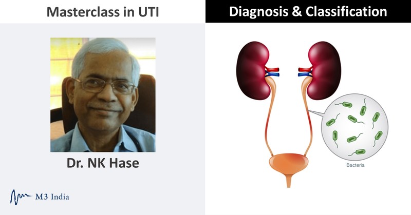
UTI diagnosis & classification with Dr NK Hase-Exclusive masterclass series -Part 1
M3 India Newsdesk Jan 09, 2022
Dr. NK Hase delivers a masterclass on urinary tract infections (UTIs), exclusive for M3 India members, in this 3-part series. In this first part, he discusses classification, pathogenesis, and microbiological spectrum of UTIs.
Definition of UTI
UTI is defined as an inflammatory response of the uroepithelium starting from the urethra to the kidney to bacterial invasion. It is usually associated with bacteriuria (bacteria in the urine) and pyuria (WBCs in the urine).
- Bacteriuria without pyuria suggests bacterial colonisation without active infection.
- Pyuria without bacteriuria is called sterile pyuria; suggests interstitial nephritis, kidney stones, and tuberculosis
Spectrum of UTI may range from asymptomatic bacteriuria to life-threatening tissue infection associated with shock and multiorgan failure.
Magnitude of problem
Urinary tract infections (UTIs) are the second-most commonly diagnosed infectious diseases worldwide in the community as well as healthcare settings. About 150 million people are diagnosed with UTI each year worldwide.
It more commonly occurs in women than in men at ratio 8:1.
- Women: Approximately 50 to 60% of women report at least one attack of UTI in their lifetime. The incidence of cystitis in young sexually active women is approximately 0.5 per person per year and recurs in 27 to 44% of healthy women with normal urinary tracts. One in every three women will have had at least one episode of UTI by the age of 24 years. Approximately 10% of post-menopausal women have UTI each year.
- Men and childen: UTI is rare and usually associated with structural and functional abnormality of urinary tract needing further investigations.
- Elderly: Incidence of UTI increases markedly in elderly. Bacteriuria is found in 21% of women and 12% of men over age of 65 years.
Eighty percent of nosocomial UTIs are secondary to indwelling urethral catheters. E.coli (Escherichia coli) is the most common pathogen in both men and women, community and healthcare settings. E. coli and Klebsiella pneumoniae are still common organisms associated with community as well as healthcare-associated UTI. ESBL-producing multidrug resistant strains are emerging making therapy of UTI challenging.
Classification of UTI
According to the site of infection UTIs are classified as upper urinary tract infections and lower urinary tract infections.
| Upper urinary tract infections | Lower urinary tract infections |
|
Kidneys: Pyelonephritis, kidney abscess, pernephric abscess General symptoms: Fever, flank pain, back pain, nausea, vomiting. costovetebral angle tenderness |
Bladder: Cystitis, urethritis, prostatitis Local symptoms: Dysuria, frequency, urgency, suprapubic pain, no systemic symptoms |
According to severity of infections, UTIs can be classified as uncomplicated or complicated UTIs. Infections can be symptomatic or asymptomatic, community-acquired or nosocomial or healthcare-associated infections.
| Uncomplicated UTI | Complicated UTI |
|
Examples: Infections in the presence of diabetes, pregnancy, stones, advance age, men, children, and neurogenic bladder. |
Pathogenesis of UTI
The human urinary tract and urine is normally sterile. There are various host factors that prevent invasion of bacteria. UTI occurs when an uropathogen from faecal flora colonises the periurethral area, enters the bladder via the urethra, adheres to the uroepithelium, and stimulates host responses. Upper tract infection most of the time occurs as an ascending infection from the lower urinary tract. And occasionally, the microorganism can reach the urinary tract by haematogenous or lymphatic spread.
Host defence factors
- Normal vaginal flora – prevent vaginal colonisation
- Lactobacilli suppress other bacteria.
- Acidic pH inhibits growth of E.coli in the vagina
- Secretary IgA in the vaginal fluid
- Prevention of bladder colonisation
- Urine flow and voiding (mechanical clearance)
- Presence of bacteriostatic substances in the urine
- Mucosal defense mechanisms
- Competent vesicourethral sphincter prevents reflux
- The ability of blood group antigen secreted into the body fluids in vaginal secretions and urine to block the adherence of bacterial adhesins to uroepithelium receptors
- In males a greater distance between anus and urethral orifice, increased urethral length and antibacterial substance in prostatic fluid acts as host defence mechanism
- Bacterial virulence factors: Uropathogenic or nephritogenic E.coli posse’s variety of virulence properties that appear to be important in mediating the key steps in pathogenesis of UTI in the normal urinary tract.
- Sustained intestinal carriage, persistence in vagina and ascension and invasion of urinary tract with normal defence mechanism
- Adhesins- Type 1 pili – promote bacterial attachment by binding to tissue surface receptors
- Filamentous bacterial appendages type 1 pili bind to mannose residue on uroepithelium
- Allow bacteria to ascend ureter
- Capsular antigen, somatic antigens promote evasion of host response
- Haemolysins aerobactin: resist bactericidal action of normal serum
Increase in virulence of uropathogen + decrease in host defence = urinary tract infections
| Host factors that decrease the host defence & predispose to urinary tract infections | |
| Young healthy women |
|
| Post-menopausal women |
|
| Urologic/structural abnormalities |
|
| Metabolic & miscellaneous |
|
| Instrumentation & urologic procedure |
|
Microbiological spectrum
- Escherichia coli (E.coli) remains the most common pathogen in 75 to 90% of uncomplicated and complicated (21 to 54%) cases of UTI.
- Staphylococcus saprophyticus is the second-most common organism responsible for UTI in young healthy women.
- Occasionally, the infection is caused by other Enterobacteriaceae species: Klebsiella pneumoniae (1 to 2%), Proteus mirabilis (1 to 2%).
- Pseudomonas, Enterococci and Staphylococcus aureus are mainly seen in nosocomial and complicated UTIs. Proteus mirabilis is frequently associated with kidney stones or structural abnormality of the urinary tract.
- Enterococci faecalis is common in the elderly; Staphylococcus aureus is usually seen with cortical renal abscesses suggesting haematogenous spread.
- Non-bacterial organisms such as fungi (Candida) commonly associated with indwelling catheters, prolonged broad spectrum antibiotic therapy and immunosuppressive therapy.
- Kidneys may be involved in systemic disseminated fungal infections such as Aspergillosis, mucor mycosis and histoplasmosis rarely in renal transplant recipients.
- Viruses like adenovirus 2, BK virus can cause haemorrhagic cystitis in immunosuppressed patients.
- Chlamydia trachomatis, Mycoplasma hominis may cause urethral syndrome.
- Isolation of organism such as lactobacilli, group B streptococci, and coagulase negative staphylococci from voided midstream urine commonly represent contamination of urine specimen.
Diagnosis of urinary tract infections
- History and physical examination may suggest site of infection
- History of dysuria, frequency, urgency, nocturia and suprapubic pain may suggest acute cystitis
- History of fever with chills flank pain abdominal pain (renal) angle tenderness with or without dysuria frequency, urgency, and nocturia may suggest pyelonephritis
Urinalysis
This is a simple, cheap, easily available method for diagnosing pyuria and bacteriuria. Mid-stream clean catch freshly voided sample is preferred. In any urine sample kept for more than 4 hours, bacteria (contaminant) grows. Unspun sample showing >10 leukocytes/mm3 is considered significant pyuria by microscopy. Spun sample showing more than 5 WBC/HPF is also considered significant but spun urine for WBC has high false positive/negative results. WBC cast if present is highly suggestive of pyelonephritis. A gram stain of unspun urine showing one or more organism per oil immersion field correlates with the presence of more than 105 bacteria/ml.
Dipstick test detects presence of leukocyte esterase (an enzyme released from leukocytes) reflecting pyuria and nitrite (reflecting the presence of Enterobacteriaceae which converts urinary nitrate to nitrite).The dipstick test is most accurate for predicting UTI when positive for both leukocyte esterase and nitrite with sensitivity of 75% and specificity of 82% and negative predictive value of 90%.This simple bedside test can diagnose UTI.
Urine culture
The cornerstone of diagnosis of UTI is a quantitative urine culture. Urine culture should be obtained before starting antimicrobial therapy. Urine specimen for culture must be collected with a method that minimises contamination. Men and women are instructed to give a mid-stream urine sample in a sterile container. Instructions to give to a patient are:
- Start with full bladder
- Stand with legs on either side of toilet
- Women- Clean vulva area with sterile swab or soapy water from front to back, hold labia apart; Men- Clean the tip of penis
- Pass urine into the toilet until half is finished
- Collect sample in a sterile pot in between, finish passing urine into the toilet
When the patient is not able to give a voided sample, urine may be collected by in and out catheter. For a patient on short-term indwelling catheter, urine sample should be collected by puncture of the catheter port. If a patient is on long-term indwelling catheter, the catheter should be removed and replaced by a new one. A specimen of bladder urine should be collected through the newly-placed catheter. Urine sample can be collected ideally by suprapubic puncture. Suprapubic puncture urine culture is considered as the gold standard for diagnosis of UTI but is not practicable.
Urine sample once collected should be transported to the laboratory for examination and culture without delay. In any urine sample kept for more than 4 hours at room temperature, bacterial growth occurs (contaminants). If the specimen is delayed, it should be refrigerated at 40ºC until transported.
| Diagnostic criteria for quantitative urine culture | |
| Asymptomatic bacteriuria (mid-stream sample) | ≥105/CFU/ml |
| Suprapubic puncture | Any growth |
| Catheter sample | ≥102/CFU/ml |
| Indwelling catheter- Asymptomatic | ≥105/CFU/ml |
| Indwelling catheter- Symptomatic | ≥102/CFU/ml |
| Women mid-stream: Symptomatic cystitis | ≥103/CFU/ml |
| Women mid-stream: Symptomatic pyelonephritis | ≥104/CFU/ml |
| Men mid-stream: Symptomatic | ≥103/CFU/ml |
Bacteriuria in the absence of pyuria is usually contamination/colonisation. Pyuria without bacteriuria suggests sterile pyuria may be due to inflammation rather than bacterial infection. Causes of sterile pyuria include interstitial nephritis stone disease, tuberculosis, uroepitheial tumour, chlamydial and mycoplasma infections.
This article was originally published on July 8, 2020.
Disclaimer- The views and opinions expressed in this article are those of the author's and do not necessarily reflect the official policy or position of M3 India.
The author Dr. NK Hase is a Director clinical Nephrology & Transplant working at Jupiter Hospital, Thane and former Professor & Head of Department of Nephrology Seth GS Medical College and KEM Hospital, Mumbai.
-
Exclusive Write-ups & Webinars by KOLs
-
 Daily Quiz by specialty
 -
 Paid Market Research Surveys
 -
Case discussions, News & Journals' summaries
