The Rare Third Leg: A Rare Case Report of Sacrococcygeal Teratoma
M3 India Newsdesk Jan 05, 2023
This article examines a unique case presentation of a 25-year-old male patient who claimed to have had a third limb from birth that ultimately grew to its current size.
Introduction
Sacrococcygeal Teratoma (SCT) is most commonly seen in neonates with a rare incidence of 1:40,000 in live births. The incidence is more common in females as compared to males with a ratio of 4:1.It is documented in the literature that they arise from totipotent somatic cells of the primitive knot. Hence they are composed of multiple tissue types comprising two or three germ layers. The presentation of this condition is very rare in adults. They are ruled out as benign in nature and have a very low potential for malignancy due to their dormant nature.
Ultrasound done in the second trimester can help in early detection. The prognosis of such cases is excellent with surgical intervention followed by coccygectomy. The consequence of not removing the coccyx may lead to recurrence ad it increases the chances of the same by 37% according to past literature.
The most common complication is the unpleasant appearance of a surgical scar. Other surgical complications could include neurogenic bladder, urinary and faecal inconsistency and other chronic problems due to the accidental damage of nerves and muscles within the pelvis. Alman et.al reported the largest set of 400 cases of SCTs and proposed the classification for the same.
A rare case-The third leg!
This is a review of a rare case report of a 25-year-old male patient, who reported the presence of a third leg since birth which eventually grew to the present size.
Patient complaints
The patient complained of difficulty in sitting, sleeping and walking and also cosmetic blemishes due to the strange appearance.
Appearance
- It was a single non-tender solid mass measuring 35 cm × 20 cm × 10 cm extending between the mid-axial lines horizontally and from the highest point of the lilac crest to the tip of the coccyx with a short 4 cm leg.
- The mass appeared like a good form foot measuring 17 cm × 8 cm with distinct heal, sole, dorsum and five toes overhanging the right buttock.
- There was evidence of rudimentary genitalia on the right side of this foot and it showed a good range of movements over its attachment at the sacrococcygeal region.
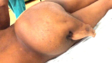
Figure 1-Posterior view-Mass extending from the highest point of the lilac crest to the tip of the coccyx with a short leg and well-formed foot overhanging the right buttock.
Radiographic appearance
- An X-ray revealed well-formed bones of the foot, especially the long bones which resemble the femur and tibia.
- A plain MRI of the lower lumbosacral region revealed an L4/L5 block vertebral anomaly of the sacrum with spina-bifida.
- Few other features like subcutaneous fat, a few rudimentary bone structure, a few muscular elements and a well-defined saccular structure showing a pocket of air, resembling a bowel, overlying the sacrum.
- There was no gross anomaly noted in the pelvis region. Ultrasound showed abnormality too. Alfa fetoprotein levels were normal.
- Provisional diagnoses given were Sacrococcygeal Teratoma-Altman Type II. In such cases, the tumour is outside the body with a small portion inside the pelvis.
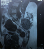
Figure 2-MRI showing a mass in the sacrococcygeal area, L4/L5 sacral anomaly with spina bifida, a few bony structures, subcutaneous fat, muscle elements and a bowel segment overlying the sacrum
Surgical intervention
The excision was planned under general anaesthesia. A vertical elliptical incision was made encircling the mass and the limb and the limb was further excised with the entire mass of muscles and bone. The location of spina-bifida was identified and the dura was safeguarded.
A triangular curved bone of 8 × 6 cm was identified near the sacrum between which a cystic mass was seen. Dissection of this bony attachment was done with an osteotome and the mass was excised. A skin-flap reconstruction was then performed and there were no intra-complications noted.
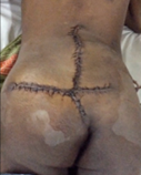
Figure 3-Post operative view of the patient with SCT excised and reconstruction performed using a flap.
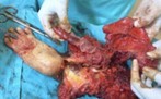
Figure 4-Exicised SCT specimen with a well-formed foot structure, subcutaneous fat and some muscle elements. bone and bowel segment
Histopathology revealed the tumour to be a mature teratoma with skin and subcutaneous fat. The excised section also showed well-differentiated bone resembling the lower limb, rudimentary testis, bowel and lymphatic tissue. There was no evidence of malignancy
Prognosis
The healing was progressive with a scar. Postoperative bowel and bladder function were normal. Both limbs showed normal motor and sensory function. A 3-year subsequent follow-up revealed no other complications.
Discussion
This condition is extremely rare in adults and including this case only eight cases have been reported and documented in the literature with an extra limb in SCT and the third one in adults. Though this condition shows a female predominance, the current case was a male.
Modern ultrasound helps in prenatal detection but as this patient was born in 1989, the diagnostic techniques were not well established. Also, the patient’s rural background and lack of awareness lead to a delayed presentation of the case and hence delayed treatment. The accessory blood supply in this case was between the rectum and perirectal muscles. In the current case, the SCT was not attached to the rectum or other important organs, hence it allowed a complete resection of the tumour after the blood supply was controlled and managed well.
Conclusion
Follow-up during the first three years is extremely important to monitor any recurrence and manage any surgical complications. In this case, no major complications were noted and no recurrence was also noted.
Disclaimer- The views and opinions expressed in this article are those of the authors and do not necessarily reflect the official policy or position of M3 India.
About the author of this article: Dr Ridima Surve is a practising dentist from Mumbai.
-
Exclusive Write-ups & Webinars by KOLs
-
 Daily Quiz by specialty
 -
 Paid Market Research Surveys
 -
Case discussions, News & Journals' summaries
