Radiation safety principles for doctors: Dr. Ravikanth Reddy
M3 India Newsdesk Mar 14, 2019
Dr. Ravikanth Reddy, a noted Radiologist stresses on the need for regulations to improve radiation safety in India and asserts that ALARA policies can be effectively implemented through understanding and using the key parameters- time, distance, and shielding.
Physicians and radiologists must be aware of the radiation risks and benefits associated with medical exposures, must be able to understand and implement the principles of radiation protection for patients in day-to-day practice. In summary, ALARA policies can be effectively implemented through understanding and using the key parameters time, distance, and shielding.
Although radiation diagnostics is an important and broadly used part of the therapeutic process, protection-related issues are usually addressed in a rather offhand manner. In the era of increasing pro-health awareness within the society as well as of increasingly common claims filed against medical personnel, a better knowledge of radiation protection issues becomes an important element of professional expertise of not only radiologists and radiation therapists, but also clinicians as well as auxiliary staff.
Diagnostic radiology procedures, such as computed tomography (CT) and X-ray, are an increasing source of ionising radiation exposure to our community.
- The knowledge of ionising radiation exposure risks among clinicians is essential for planning diagnostic procedures and therapy. The common access to imaging methods based on ionising radiation also requires radiation protection.
- Exposure to ionising radiation is associated with increased risk of malignancy, proportional to the level of exposure. Every diagnostic test using ionising radiation needs to be justified by clinical need.
- Clinicians need a working knowledge of radiation safety so they can adequately inform their patients of the risks and benefits of diagnostic imaging procedures.
The increased use of ionisation radiation for diagnostic and therapeutic purposes, the rapid advances in computed tomography as well as the high radiation doses delivered by interventional procedures have raised serious safety and health concerns for both patients and medical staff and have necessitated the establishment of a radiation protection committee (RPC) in every Radiology Department. To ensure the safety of patients, providers, and staff members, it is important that the health care community becomes familiar with the terminology, common equipment, and standard practices used in radiation safety and monitoring.
Further, Dr. Reddy makes an attempt to provide an introduction to radiation safety and monitoring so that health care providers and support personnel who may have contact with patients, equipment, and facilities that use radiation / radioactive material may be more aware of the policies and precautions that are in place to ensure their safety.
Basics of Radiation Safety
In radiation protection, the guiding philosophy is ALARA (as low as reasonably achievable), and states have regulatory authority. Dose limits are in part based on effective dose equivalent and differences in tissue sensitivities. Interventional cardiac procedures represent the largest contribution of ionising radiation source after computerised tomography and nuclear medicine. Modern therapies and the need for quality radiological imaging have dramatically increased the use of ionizing radiological imaging in cardiology.
In diagnostic radiology, the main source of occupational dose is scattered radiation from the patient--particularly from fluoroscopically guided procedures. Personnel stand near patients for long times, and angulated geometries with C-arm equipment may result in high personnel doses from backscatter. Although dose limits typically regulate maximum whole-body dose, protective clothing worn by fluoroscopists reduces personnel risks; weighting factors can be applied to estimate effective dose equivalent.
Quantities and units for quantifying ionising radiation
Absorbed Dose: A useful quantity in radiation physics is the energy actually deposited in a certain amount (mass) of tissue. This unit is referred to as absorbed dose. The unit of absorbed dose is the gray (Gy), formerly the rad; the gray is equivalent to the absorption of one Joule of energy per kilogram. One gray equals 100 rad.
Quantities and units for quantifying biological risks
Equivalent Dose: The dosimetric quantity that accounts for the differences in biological effectiveness of various types of radiation and that allows doses from different radiations to be combined is called the equivalent dose.
The traditional unit for dose equivalent is the rem, and although this unit has been replaced by the sievert (1 Sv = 100 rem), it is still used in radiation protection regulations, and dosimetry reports present data in units of millirem. It is calculated by multiplying the absorbed dose by the appropriate radiation weighting factor, "wR".
Biological effects of radiation
- Deterministic effects: Here, an identifiable threshold level exists and the severity of effect intensifies with increasing dosage of exposure. Biological effects occur as a result of cell damage and death. Symptoms are related to the extent of cell death. Dermatological effects and cataracts are typical examples of deterministic effects.
- Stochastic effects: These follow a linear non-threshold theory, which essentially means these effects occur by chance. There is no minimum exposure, and risk increases linearly with radiation dose received. Cancer in an exposed individual occurs due to the mutation of cells as a result of chromosomal translocations.
Categories of exposure
- Occupational exposure: It is defined as all radiation exposure of workers incurred as a result of their work. International Committee on Radiology Protection (ICRP) limits its use of ‘occupational exposures’ to radiation exposures incurred at work as a result of situations that can reasonably be regarded as being the responsibility of the operating management.
- Public exposure: It encompasses all exposures of the public other than occupational exposures and medical exposures of patients. It is incurred as a result of a range of radiation sources. The component of public exposure due to natural sources is by far the largest, but this provides no justification for reducing the attention paid to smaller, but more readily controllable, exposures from man-made sources.
- Medical exposure of patients: Radiation exposures of patients occur in diagnostic, interventional, and therapeutic procedures. There are several features of radiological practices in medicine that require an approach that differs from the radiological protection in other planned exposure situations.
Relative comparison of effective doses in radiological exposures
| Type of exposure | Effective dose (mSv) | Equivalent to days of background radiation |
| Airplane | .01 to .03 | 1 to 4 days |
| Chest X-ray | .1 | 10 days |
| Mammogram | .7 | 3 months |
| CT Scan: Head | 2 | 8 months |
| CT Scan: Chest | 8 | 2 years |
| CT Scan: Pelvis | 10 to 20 | 3 to 6 years |
| MiniCAT CT Scan: Head | .17 | 20 days |
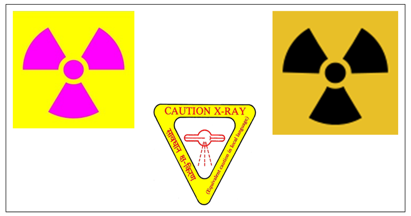
Radiation symbols
To make everybody aware of a hazard in any area where there exists a source of radiation and one can get exposed to it or may get contaminated and should take precautionary measures to avoid it, generally the trefoil ionizing radiation symbols are used.
Unique characteristics of radiological protection in medicine
The exposure of patients is deliberate. Except in radiation therapy, it is not the aim to deliver radiation dose, but rather to use the radiation to provide diagnostic information or to conduct an interventional procedure. The voluntary decision is made with varying degrees of informed consent that includes not only the expected benefit but also the potential risks.
The amount of information provided in order to obtain informed consent varies based on the exposure level (whether diagnostic, interventional, or therapeutic) and the possible emergent medical circumstances that may be attributable to radiation exposure.
Special considerations
Children and radiation exposure
Large-scale quality improvement to promote radiation protection for children is being aggressively pursued by numerous international organizations. These international agencies use quality improvement methods on a global scale to optimize medical imaging for all diagnostic imaging modalities that use ionizing radiation with the intent of lowering radiation dose to children.
Pregnant patient and radiation exposure
Radiation doses have been studied and are well known for typical diagnostic examinations. Therefore, in patients who are pregnant or potentially pregnant, it is imperative that special considerations be given. Most diagnostic x-ray studies that are performed looking at structures other than the pelvis, ovaries, uterus, and lumbar spine do not lead to measurable exposure of radiation to the foetus.
Those studies that have a higher risk of exposure include x-rays of the lumbar spine, intravenous pyelogram, and upper and lower gastrointestinal series; studies of the gallbladder and gallbladder function, pelvic, hip, and abdominal X-rays; and specific x-rays of the uterus and fallopian tubes, such as a hysterosalpingogram (HSG).
Pregnant personnel have lower limits, which apply only with voluntary declaration of pregnancy. With appropriate precautions, fetal doses can typically remain within recommended limits without changes in occupational tasks. Radiation workers in every state must ensure that regulations are appropriate.
Radiation health hazards
- Posterior sub-capsular cataracts have been reported in 50% of cardiologists and 41% of nurses working in interventional catheterization laboratories.
- Several case reports of brain tumors have emerged in the literature.
- Others include thyroid changes, neoplasm, hypertension, hyperlipidaemia, reproductive and even psychological effects have been described. Hair loss and skin damage may follow prolonged exposure during fluoroscopic procedures. These vary from temporary erythema to necrosis of the skin and subcutaneous tissues.
Radiation safety policies
Personnel dosimeter policies need to be in place for all occupationally exposed individuals, regardless of pregnancy. All employers are required to provide a dosimeter to all employees who have a likelihood of receiving doses as large as 10% of the applicable legal limit (whole body, lens of the eye, extremity). All personnel who perform x-ray procedures can potentially receive doses sufficiently large and require at least one dosimeter for monitoring whole-body dose.
Radiation safety philosophy and ALARA
ALARA represents a practice mandate adhering to the principle of keeping radiation doses to patients and personnel As Low As Reasonably Achievable (ALARA). This concept is strongly endorsed by the Society for Pediatric Radiology, particularly in the use of procedures and modalities involving higher radiation doses such as CT and fluoroscopic examinations of pediatric patients.
There is no doubt that medical imaging, which has undergone tremendous technological advances in recent decades, is integral to patient care. Current imaging methods must be optimized for radiation dose reduction in pediatric patients who might be as much as ten times more radiosensitive than adults.
General priniciples of radiation protection based on ICRP recommendation
- The principle of justification: Any decision that alters the radiation exposure situation should do more good than harm. This means that, by introducing a new radiation source, by reducing existing exposure, or by reducing the risk of potential exposure, one should achieve sufficient individual or societal benefit to offset the detriment it causes.
- The principle of optimization of protection: The likelihood of incurring exposures, the number of people exposed, and the magnitude of their individual doses should all be kept as low as reasonably achievable (ALARA), taking into account economic and societal factors. This means that the level of protection should be the best under the prevailing circumstances, maximizing the margin of benefit over harm.
- The principle of dose limitation: The total dose to any individual from regulated sources in planned exposure situations other than medical exposure of patients should not exceed the appropriate limits recommended by the International Commission on Radiological Protection (ICRP).
Radiation safety regulations in India
- No person shall, without a license:
- establish a radiation installation for design, construction, commissioning and operation
- decommission a radiation installation
- No person shall handle any radioactive material, or operate any radiation generating equipment except in accordance with the terms and conditions of a license.
- A license shall be issued for sources and practices associated with the operation of:
- nuclear fuel cycle facilities
- land based high intensity gamma irradiators other than gamma irradiation chambers
- particle accelerators used for research and industrial applications
- neutron generators
- facilities engaged in the commercial production of radioactive material or radiation generating equipment
- telegamma and accelerators used in radiotherapy
- computed tomography (CT) unit
- interventional radiological x-ray unit
- industrial radiography
- such other source or practice as may be notified by the competent authority, from time to time
Radiological Safety Officer (RSO)
Every employer shall designate, with the written approval of the competent authority, a person having appropriate qualifications as Radiological Safety Officer (RSO). Responsibilities of the Radiological Safety Officer include:
- To carry out routine measurements and analysis on radiation and radioactivity levels in the controlled area, supervised area of the radiation installation and maintain records of the results thereof.
- To investigate any situation that could lead to potential exposures; ensure that,
- reports on all hazardous situations along with details of any immediate remedial actions taken are made available to the employer and licensee for reporting to the competent authority and a copy endorsed to the competent authority
- quality assurance tests of structures, systems, components and sources, as applicable are conducted; and (iii) monitoring instruments are calibrated periodically
- To assist the employer in,
- instructing the workers on hazards of radiation and on suitable safety measures and work practices aimed at optimizing exposures to radiation sources
- the safe disposal of radioactive wastes
- developing suitable emergency response plans to deal with accidents and maintaining emergency preparedness
Radiation Safety Committee (RSC)
The establishment of a RSC requires continuing education of the staff and professional, effective communication among stakeholders of all levels and implementation of quality assurance programs. The RSC creation is being driven from the highest level.
Management, professionals and related authorities are recognised to play a vital role in the embedding and promotion of RPC in a Hospital. The combination of knowledge, values, behaviours and experience of radiation protection in all its aspects for patients, workers, population and environment, and in all exposure situations.
The objectives of RSC are:
- to provide a safe working environment
- promote knowledge of radiation risks
- minimise unsafe practices
- control radiation risks
- share responsibility among workers and improve the quality of an already existing radiation protection program
All the aforementioned objectives are achieved through the active participation and interaction of all the workers inside the radiology department. The establishment of a RSC enables the reduction of the radiation dose, enhances radiation risk awareness, minimizes unsafe practices, and improves the quality of a radiation protection program.
Establishing a Radiation Safety Program (RSP)
The most important factor contributing to the creation of a RSP is the continuous education and training of the staff and professionals with the attendance of courses, workshops, seminars and electronic-learning programs in a normal periodical basis.
Theoretical education and practical training in radiation protection aim to ensure that healthcare professionals will obtain a strong foundation in radiation protection and a basic knowledge of the technology of each modality. Education is an essential aspect for the optimisation of clinical protocols and the reduction of radiation exposure.
Clinicians, radiographers, nurses and other medical staff need to have a substantial knowledge of radiation protection regulations and a comprehensive understanding of the factors that affect patient and occupational dose in order to minimize the harmful effects of ionizing radiation.
Quality assurance (QA) program is another essential step in the implementation of RSP. According to WHO, the QA program in diagnostic radiology is defined as “an organized effort by the staff operating a facility to ensure that the diagnostic images produced are of sufficiently high quality so that they consistently provide adequate diagnostic information at the lowest possible cost and with the least possible exposure of the patient to radiation”.
The objectives of a QA program is to improve patient care and comfort, ensure accurate diagnosis and proper function of the equipment, produce high quality images following the ALARA principle, ensure patient and staff safety and minimise cost.
Impact of Radiation Safety Programs (RSP)
The direct impact of the implementation of RSP is the substantial reduction of radiation dose on both patients and staff. A strong RSP enables more efficient diagnosis and treatment and helps minimize harmful effects. The foundation of a RSP has an important effect on the operation of the Radiology Department. It improves the efficiency and service quality, reduces cost and incorrect practices and promotes a good reputation.
Current status of medical radiation exposure
Multi-detector CT, interventional procedures in radiology and cardiology contribute heavily to diagnostic radiation exposures in the current time. The significant change in ionising radiation exposure is due to an increase in medical exposures and have increased significantly in recent times.
Basic training should be available for all healthcare workers in the catheterization laboratory, and ongoing radiation safety courses should be obligatory. Unless training units actively promote and examine fellows on radiation safety, little will change.
Current trends of radiation safety protocols in India
Atomic Energy Regulatory Board (AERB), Government of India operates a management program that includes the following:
- Radiological Policy Statements
- Radiological Control Procedures
- Individual Radiological Doses
- Internal and External Dosimetry Policies and Procedures
- Personnel Training
- As Low as Reasonably Achievable (ALARA) Records
- Radiological Instrumentation Test, Repair and Calibration Records
- Radiological Surveys
- Area Monitoring Dosimetry Results
- Radiological Work Permits
- Radiological Performance Indicators and Assessments
- Radiological Safety Analysis and Evaluation Reports
- Quality Assurance Records
- Radiological Incident and Occurrence Reports
- Accountability Records for Sealed Radioactive Sources
- Records for Release of Material to Controlled Areas
- Reports of Loss of Radioactive Material
Disclaimer- The views and opinions expressed in this article are those of the author's and do not necessarily reflect the official policy or position of M3 India.
-
Exclusive Write-ups & Webinars by KOLs
-
 Daily Quiz by specialty
 -
 Paid Market Research Surveys
 -
Case discussions, News & Journals' summaries
