SFU engineer develops retina scanner that will diagnose eye diseases before vision loss occurs
Simon Fraser University News Dec 22, 2017
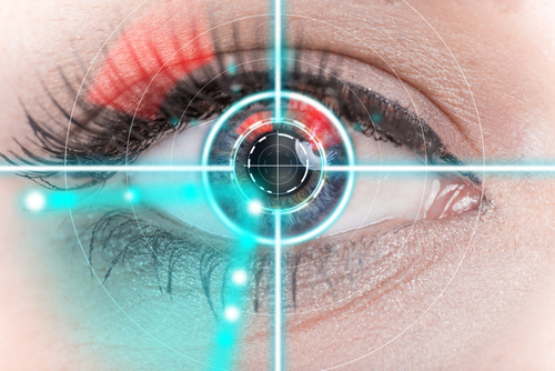
SFU engineering science professor Marinko Sarunic has developed a high-resolution retinal imaging scanner that will one day revolutionize eye care, helping ophthalmologists diagnose eye diseases before vision loss occurs.
The retina is the light-sensitive tissue at the back of the eye. Its 100 million photoreceptors convert light into the images that our brain ‘sees.’
Today, there are approximately one million Canadians with vision loss caused by major eye diseases such as wet age-related macular degeneration (Wet AMD), diabetic retinopathy, glaucoma, and others. The prevalence of vision loss in Canada is expected to double in the next 25 years. An estimated 75% of vision loss can be treated or prevented through early detection.
Sarunic’s high-resolution scanner is on the cutting edge of vision science because it can produce high-resolution, 3-D cross-sectional images of the retina—including individual photoreceptors, and fine capillaries, or blood vessels. And unlike other high-resolution retinal scanners, which are the size of a billiard table, Sarunic’s is the size of a shoebox. It’s perfect for everyday use in medical clinics and hospitals.
“It’s a breakthrough in clinical diagnostics,” says Sarunic. “With the high-resolution scanner, ophthalmologists and optometrists can detect damage and changes to small numbers of individual photoreceptors, giving them a diagnosis before the patient loses vision, and the potential to take preventative measures.”
Currently, physicians use low-resolution scanners that can only assess and diagnose the cause of dead retina cells after a patient’s vision is impacted. Last year, ophthalmologists at Vancouver General Hospital’s (VGH) Eye Care Centre spent 8 months testing Sarunic’s high-resolution scanner.
Dr. Eduardo Navajas, a vitreoretinal specialist, says the scanner eliminates the need for, and the complications related to, dye injections that are currently used to diagnose and monitor eye diseases like diabetic retinopathy and Wet AMD.
“Early detection of abnormal blood vessels caused by Wet AMD and diabetes is essential to saving a patient’s vision,” says Navajas. “Dr. Sarunic’s new imaging technology is benefiting patients, allowing us to diagnose and treat Wet AMD and diabetic eye disease before patients develop bleeding and permanent damage to their retina.”
-
Exclusive Write-ups & Webinars by KOLs
-
 Daily Quiz by specialty
 -
 Paid Market Research Surveys
 -
Case discussions, News & Journals' summaries
